1.1 - World of Microorganisms
Microorganisms and Normal Flora
- Microorganisms are minute organisms that cannot be seen with the naked eye; they can only be seen with the help of a microscope.
- Normal flora refers to microorganisms found in organisms, including humans and animals, which do not cause illness.
Normal flora in humans
- Skin: Staphylococcus sp. and Corynebacterium sp.
- Upper respiratory tract: Staphylococcus sp. and Streptococcus sp.
- Small intestine: Escherichia coli, Lactobacillus sp. and Streptococcus sp.
- Urethra: Staphylococcus sp. and Corynebacterium sp.
Importance of normal flora to human health
- Competes with pathogens for nutrients and prevents the formation of pathogen colonies.
- Includes bacteria that synthesise vitamin B₁₂ and vitamin K.
- Stimulates the growth of body tissues such as colon tissue and tissues in the digestive tract.
- Stimulates the production of antibodies that fight pathogens and diseases.
Classification of Microorganisms
Microorganisms are classified into fungi, algae, protozoa, bacteria and viruses according to their size, shape or basic structure, nutrition, habitat and method of reproduction.
Fungi
| Characteristic | Details |
|---|---|
| Size | Macroscopic fungi such as mushrooms can be seen with the naked eye. Microscopic fungi such as yeast and mucor measure 10 μm–100 μm and require a microscope. Mucor normally exists in colonies. |
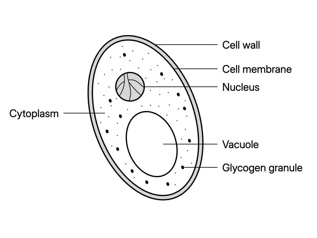
| Shape / structure | Unicellular yeast is shaped like a small sphere. Multicellular mucor consists mainly of spherical sporangia and filamentous hyphae. A yeast cell has a cell wall, cell membrane, cytoplasm, nucleus, vacuole and glycogen granules. |
| Nutrition | Saprophytic fungi obtain nutrients from dead and decaying organisms; parasitic fungi obtain nutrients from their hosts. |
| Habitat | Places rich in decaying matter, faeces, animal skin and food; fungi grow well in dark, moist places. |
| Reproduction | Yeast: asexual budding. Mucor: asexual spore formation or sexual conjugation. |
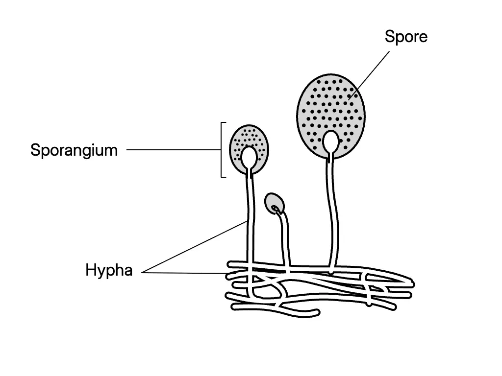
Spore formation in mucor: sporangium releases microscopic, fine and light spores → wind carries spores to a favourable environment → spores germinate without fertilisation.
Conjugation in mucor: hyphae meet → gametes form → fertilisation of gametes produces new mucor.
Algae
| Characteristic | Details |
|---|---|
| Size | Macroscopic sea algae can be seen with the naked eye. Microscopic Chlamydomonas sp. and Spirogyra sp. measure from 1 μm to hundreds of μm. |
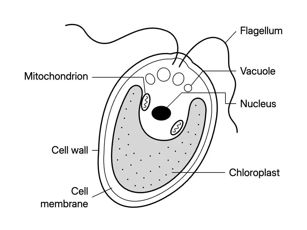
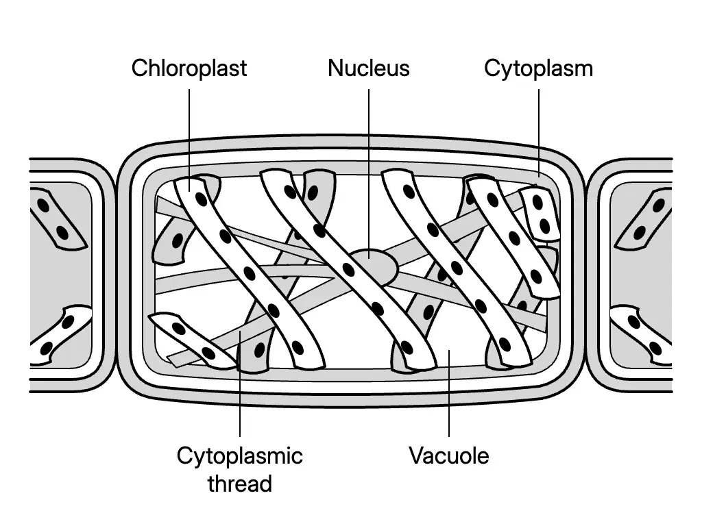
| Shape / structure | Unicellular Chlamydomonas sp. and multicellular Spirogyra sp. have chloroplasts containing chlorophyll. Chlamydomonas sp. also has flagella; Spirogyra sp. has cytoplasmic threads. |
| Nutrition | Most algae contain chlorophyll and carry out photosynthesis to produce their own food. |
| Habitat | Fresh water, salt water, moist soil and tree bark exposed to sunlight. |
| Reproduction | Chlamydomonas sp.: asexual binary fission and sexual conjugation. Spirogyra sp.: sexual conjugation. |
Protozoa
| Characteristic | Details |
|---|---|
| Size | Mostly unicellular, measuring 5 μm–250 μm; visible under the low powers of a light microscope. Protozoa normally exist in colonies. |
| Shape / structure | Paramecium sp. is slipper-shaped and has a micronucleus, macronucleus, cytoplasm, food vacuoles, contractile vacuoles, cell membrane and cilia. Amoeba sp. has no fixed shape and has a nucleus, cytoplasm, food vacuoles, contractile vacuoles and cell membrane. |
| Nutrition | Euglena sp. carries out photosynthesis. Plasmodium sp. is parasitic. Amoeba sp. obtains nutrients by phagocytosis. |
| Habitat | Paramecium sp.: fresh water. Amoeba sp.: moist soil, fresh water, seawater and its host. |
| Reproduction | Paramecium sp. and Amoeba sp.: asexual binary fission. Paramecium sp. also reproduces sexually by conjugation. |
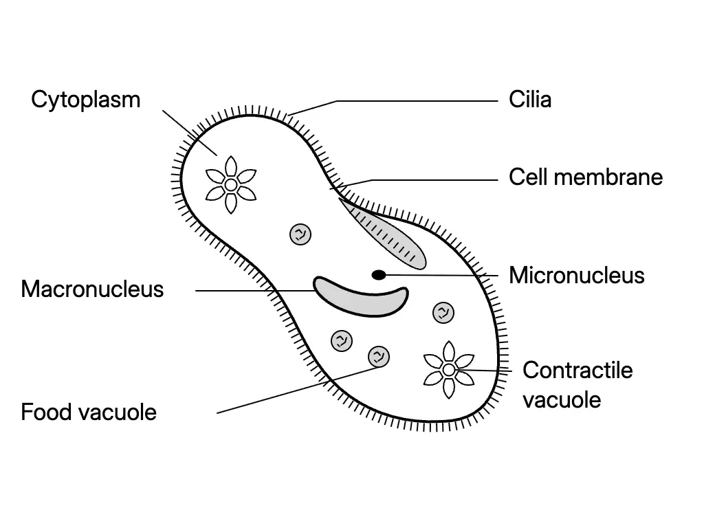
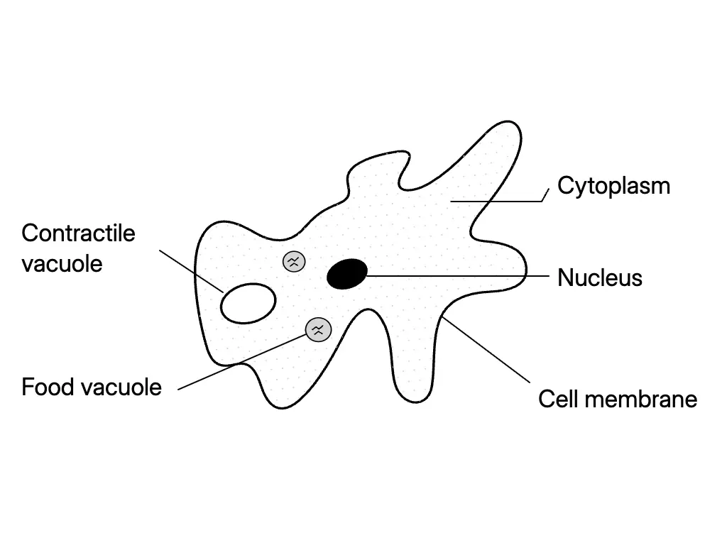
Phagocytosis in Amoeba sp.: moves towards food → pseudopodia (“false feet”) surround and engulf food → food vacuole forms → food is digested and nutrients are absorbed.
Binary fission: division of nucleus → division of cytoplasm → two daughter cells with genetic material similar to the parent.
Conjugation in Paramecium sp.: two cells unite and exchange genetic materials.
Bacteria
| Characteristic | Details |
|---|---|
| Size | Unicellular microorganisms measuring 0.2 μm–10 μm; visible under the high powers of a light microscope. |
| Basic shapes | Spherical (coccus), spiral (spirillum), rod (bacillus) and comma (vibrio). Examples: Streptococcus sp., Treponema pallidum, Bacillus anthracis and Vibrio cholerae respectively. |
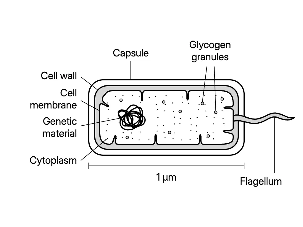
| Basic structure | Cell wall, cell membrane, cytoplasm, genetic material and glycogen granules. Some have a protective capsule, pili for attachment or a flagellum for movement. |
| Nutrition | Bacteria with chlorophyll make their own food. Parasitic bacteria obtain nutrients from a host. Saprophytic bacteria obtain nutrients from dead organisms. |
| Habitat | Air, water, soil, and all organisms and decaying materials. |
| Reproduction | Asexual binary fission and sexual conjugation. |
- Most bacterial cell walls are firm, give shape and support, and are made of amino acids and polysaccharides, not cellulose.
- During bacterial conjugation, a conjugation tube forms between donor and recipient cells; one strand of a plasmid is transferred. A plasmid resembles a small circular chromosome, can replicate independently and transfers genetic material.
- Bacteria such as Bacillus anthracis form endospores to withstand extreme heat or cold, drought and food shortage.
- An endospore forms inside a bacterial cell and has a strong protective coat. Under favourable conditions, the coat ruptures and the endospore germinates into a new bacterium.
Viruses
| Characteristic | Details |
|---|---|
| Size | The smallest microorganisms, measuring less than 0.5 μm; visible only under an electron microscope. |
| Shape | Spherical, helix, polyhedral or complex (a combination of helix and polyhedral). Examples: influenza virus, mosaic virus, adenovirus and bacteriophage. |
| Basic structure | A nucleic acid thread—DNA or RNA—protected by a protein layer called a capsid. The capsid determines the virus shape. |
| Special characteristic | Outside a host, viruses do not respire, excrete, grow or respond to stimuli. They reproduce only by infecting host cells such as bacterial, animal or plant cells. |
Reproduction of a bacteriophage in a bacterial cell:
- The virus attaches to the surface of the bacterial host cell.
- It injects its genetic material, DNA, into the bacterial cell.
- Viral genetic material controls the bacterial cell and uses its materials to build capsids.
- The number of new viruses inside the bacterial cell increases.
- New viruses rupture the cell, are released and infect other bacterial cells.
Experiment 1.1 — Presence of Microorganisms
Investigation framework
- Aim: Compare bacterial growth on sterile nutrient agar streaked with unwashed fingers, fingers washed with water only, and fingers washed with soap and water.
- Problem statement: How does finger cleanliness affect the rate of bacterial growth on sterile nutrient agar?
- Hypothesis: As finger cleanliness increases, bacterial growth decreases.
- Manipulated variable: Cleanliness of fingers used to streak the agar.
- Responding variable: Number of bacterial colonies on the agar.
- Constant variable: Surrounding temperature.
- Materials: Sterile nutrient agar, cellophane tape and marker pen.
- Apparatus: Four sterile Petri dishes with lids (A–D) and a sterile 10 cm³ measuring cylinder.
Method and expected observation
- Place 10 cm³ sterile nutrient agar in each Petri dish.
- Streak A with unwashed fingers; wash hands with water only and streak B; wash hands with soap and water and streak C.
- Leave D unstreaked as the control set-up.
- Cover, seal with cellophane tape and invert all dishes.
- Keep at room temperature in a dark cupboard for three days; compare colony numbers.
| Petri dish | Expected relative number of bacterial colonies |
|---|---|
| A: unwashed fingers | Most |
| B: fingers washed with water only | Fewer than A |
| C: fingers washed with soap and water | Fewest among A–C |
| D: not streaked | None if sterility is maintained |
- Conclusion: Washing with soap and water removes more bacteria than washing with water only; microorganisms are present on unwashed fingers.
- Sterile agar and Petri dishes ensure that observed colonies originate from the tested fingers, not pre-existing contamination.
- Sealing prevents entry or escape of microorganisms; inversion prevents condensed water from dripping onto the agar; darkness promotes bacterial growth.
- The sterile agar in D verifies that the agar and dish were initially free of microorganisms.
Safety precautions for microorganism experiments
- Wash hands with soap and water before and after the experiment.
- Wear gloves when handling bacterial cultures.
- Keep Petri dishes sealed after inoculation; do not open cultured dishes.
- Sterilise all waste before disposal.
- Immerse used apparatus in disinfectant after the experiment.
Factors Affecting the Growth of Microorganisms
| Factor | Condition favouring growth / comparison |
|---|---|
| Nutrition | Nutrient-rich conditions promote growth; compare nutrient agar with non-nutrient agar. |
| Humidity | High humidity promotes growth; compare moist nutrient agar with dry nutrient agar. |
| Light | Dark conditions promote the growth of Bacillus sp.; compare darkness with a bright area. |
| Temperature | Bacillus sp. grows best at room temperature in the stated investigation; compare room temperature, 5°C and 70°C. |
| pH value | Bacillus sp. grows most rapidly at pH 7; compare neutral, acidic and alkaline media. |
Common inoculation technique for Experiment 1.2
- Sterilise a wire loop in a Bunsen burner flame until it glows; cool it to room temperature.
- Dip the sterile loop into Bacillus sp. culture solution and smear the culture in a zigzag pattern over the agar.
- Cover and seal the Petri dish with cellophane tape, then invert it.
- Maintain the stated conditions for three days.
- Record the number of colonies as none, few or many. More colonies indicate greater bacterial growth.
Experiment 1.2A — Effect of nutrition
- Hypothesis: Bacillus sp. grows more rapidly when nutrients are present.
- Variables: manipulated—presence of nutrients; responding—number of colonies; constant—volume of culture solution and surrounding temperature.
- Set-ups: A contains 10 cm³ sterile nutrient agar; B contains 10 cm³ sterile non-nutrient agar. Inoculate both and keep inverted at room temperature in a dark cupboard for three days.
- Expected result / conclusion: A has more colonies than B; nutrients promote growth.
Experiment 1.2B — Effect of humidity
- Hypothesis: Low humidity retards the growth of Bacillus sp.
- Variables: manipulated—moisture content of nutrient agar; responding—number of colonies; constant—volume of culture solution and surrounding temperature.
- Set-ups: C contains moist sterile nutrient agar. Heat D in an oven until its nutrient agar is dry, then cool it to room temperature before inoculation. Keep both inverted at room temperature in a dark cupboard for three days.
- Expected result / conclusion: C has more colonies than D; high humidity promotes bacterial growth.
- Cooling D before inoculation prevents heat from killing bacteria and keeps temperature from becoming an unintended variable.
Experiment 1.2C — Effect of light
- Hypothesis: Light retards the growth of Bacillus sp.
- Variables: manipulated—presence of light; responding—number of colonies; constant—volume of culture solution and surrounding temperature.
- Set-ups: Keep inoculated E inverted in a dark cupboard and inoculated F inverted in a bright area near a window for three days.
- Expected result / conclusion: E has more colonies than F; darkness promotes bacterial growth.
- F is not placed under direct sunlight because sunlight would change its temperature, preventing a fair test of light alone.
Experiment 1.2D — Effect of temperature
- Hypothesis: Growth is highest at room temperature.
- Variables: manipulated—temperature; responding—number of colonies; constant—volume of culture solution.
- Set-ups: Keep inoculated G at room temperature in a dark cupboard, H in a refrigerator at 5°C, and I in an incubator at 70°C, all inverted, for three days.
- Expected result / conclusion: G has the most colonies; low temperature retards growth, very high temperature kills bacteria or retards growth, and room temperature is most favourable among the tested temperatures.
Experiment 1.2E — Effect of pH value
- Hypothesis: Growth is most rapid at pH 7.
- Variables: manipulated—pH value; responding—number of colonies; constant—volume of culture solution and surrounding temperature.
- Set-ups: Mix 10 cm³ sterile nutrient agar with 1 cm³ distilled water in J (pH 7), 1 cm³ dilute hydrochloric acid in K (pH below 7), and 1 cm³ dilute sodium hydroxide solution in L (pH above 7). Inoculate, invert and keep in a dark cupboard at room temperature for three days.
- Expected result / conclusion: J has the most colonies; neutral conditions at pH 7 are most favourable to Bacillus sp., while acidic and alkaline conditions retard growth.
- Daily-life application: Acidic food preservation methods, such as pickling, retard bacterial growth by lowering pH.
Answer practice questions to test your knowledge
Practice