Transport in Humans and Animals
10.2 - Circulatory System of Humans
Components of the Human Circulatory System
- Blood: Connective tissue made up of blood plasma, blood cells and platelets; acts as a transportation medium.
- Heart: Muscular pump that circulates blood throughout the body.
- Blood vessels: Arteries, capillaries and veins connected to the heart; transport blood to body tissues.
Structure of the Human Heart and Associated Blood Vessels
Chambers and Walls
- The heart lies between the lungs in the thoracic cavity.
- Four chambers: left atrium, right atrium, left ventricle and right ventricle.
- Atria receive blood returning to the heart; ventricles pump blood out.
- Ventricular walls are thicker and contract more strongly than atrial walls.
- The left ventricle wall is thicker than the right because it generates greater pressure to pump blood through the aorta to the whole body; the right ventricle pumps only to the lungs.
- The septum separates the left and right sides and prevents oxygenated blood from mixing with deoxygenated blood.
Valves of the Heart
- Tricuspid valve: Between the right atrium and right ventricle; three leaflets; prevents backflow into the right atrium.
- Bicuspid valve: Between the left atrium and left ventricle; two leaflets; prevents backflow into the left atrium.
- Semilunar valves: At the bases of the aorta and pulmonary artery; prevent backflow into the ventricles when they relax.
Main Blood Vessels Associated with the Heart
- Aorta: Main artery; transports oxygenated blood to the whole body.
- Vena cava: Main vein; transports deoxygenated blood back to the heart.
- Pulmonary artery: Transports deoxygenated blood from the heart to the lungs.
- Pulmonary veins: Transport oxygenated blood from the lungs to the heart.
- Coronary arteries: Transport oxygenated blood to heart tissues.
- Coronary veins: Transport deoxygenated blood from heart tissues.
Comparison of Blood Vessels: Arteries, Capillaries and Veins
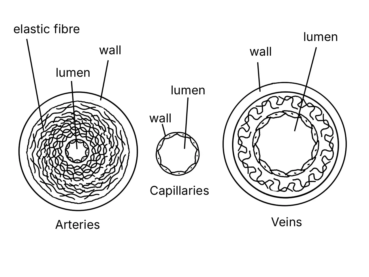
Structural and Functional Differences
- Arteries:
- Transport blood away from the heart rapidly and at high pressure.
- Thick, muscular, elastic wall; small lumen.
- No valves except semilunar valves at the bases of the aorta and pulmonary artery.
- Branch into arterioles, which end in capillary networks.
- Blood capillaries:
- Wall is one cell thick, non-muscular and non-elastic; lumen is very small.
- Allow gases, nutrients, excretory substances and hormones to diffuse between blood and cells.
- Join to form venules; flow is from arteries to veins at low pressure.
- Veins:
- Transport blood from the body back to the heart at very low pressure.
- Thin, less muscular, less elastic wall; large lumen.
- Valves maintain one-way blood flow.
Composition of Human Blood
Blood Plasma (55% of Blood Volume)
- Water (90% of plasma): Transportation medium and solvent for respiratory gases, ions, digestive products and excretory substances.
- Fibrinogen: Blood clotting.
- Albumin: Controls blood osmotic pressure.
- Globulin: Antibody involved in body defence.
- Solutes: Nutrients such as glucose, excretory substances such as urea, and respiratory gases.
- Hormones: Control physiological activities.
- Enzymes: Involved in cellular metabolic processes.
Cell Components (45% of Blood Volume)
- Erythrocytes (red blood cells):
- Elastic plasma membrane; biconcave disc shape provides a large TSA/V for efficient gaseous exchange.
- No nucleus when mature, allowing more haemoglobin to be loaded.
- Produced in bone marrow such as the sternum and ribs; live up to 120 days; destroyed in the liver or spleen by phagocytosis.
- Haemoglobin contains an iron atom that binds oxygen; it forms oxyhaemoglobin at high oxygen partial pressure and releases oxygen at low oxygen partial pressure.
- Platelets:
- Fragments of cell cytoplasm originating from bone marrow; lifespan of about one week.
- Involved in blood clotting.
- Leucocytes (white blood cells):
- Irregular shape; nucleus present; no haemoglobin; produced in bone marrow; lifespan under five days.
- Diffuse through capillary pores to fight pathogens in tissue fluid.
- Granulocytes: neutrophils engulf bacteria and dead cells by phagocytosis; eosinophils release enzymes that fight inflammation and allergic reactions; basophils contain heparin that prevents blood clotting.
- Agranulocytes: lymphocytes produce antibodies and antitoxins; monocytes engulf bacteria and dead cells by phagocytosis.
Answer practice questions to test your knowledge
Practice10.1 - Types of Circulatory System10.2 - Circulatory System of Humans10.3 - Mechanism of Heartbeat10.4 - Mechanism of Blood Clotting10.5 - Blood Groups of Humans10.6 - Health Issues Related to the Human Circulatory System10.7 - Lymphatic System of Humans10.8 - Health Issues Related to the Human Lymphatic System