Homeostasis and the Human Urinary System
13.2 - The Urinary System
Structure and Functions of the Kidney
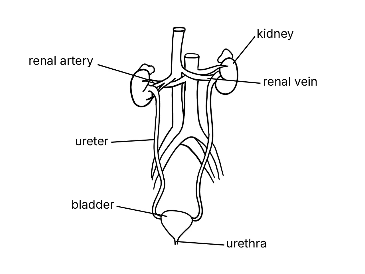
- The urinary system consists of the kidneys, ureters, bladder and urethra.
- It excretes nitrogenous compound wastes such as urea.
- It regulates body-fluid volume, blood osmotic pressure, ion concentration in body fluids, electrolyte content and blood pH.
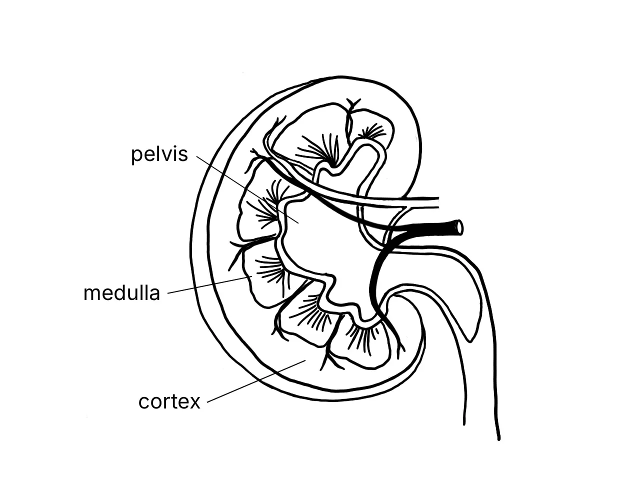
- The kidneys consist of the cortex and medulla; urine formed in the kidneys flows into the pelvis.
Main Functions of the Kidneys
- Excretion: Excretes toxic nitrogenous wastes such as urea, uric acid, ammonia and creatinine.
- Osmoregulation: Controls the total volume of water and ion concentration in body fluids, blood osmotic pressure, electrolyte content, and the pH of blood and body fluids.
Blood Supply and Urine Flow
- The renal artery carries oxygenated blood from the heart to the kidneys.
- The renal vein carries deoxygenated blood from the kidneys back to the heart.
- Urine flows through the ureter to the bladder.
Structure of the Nephron and Collecting Duct
- Each kidney contains millions of functional units called nephrons.
- Each nephron consists of the Bowman’s capsule, glomerulus and renal tubule.
- The cup-shaped Bowman’s capsule contains a cluster of blood capillaries called the glomerulus.
- The glomerulus is formed from the afferent arteriole branching from the renal artery; the capillaries merge to form the efferent arteriole.
- The renal tubule consists of the proximal convoluted tubule, loop of Henle and distal convoluted tubule.
- The loop of Henle is a long U-shaped tubule extending into the renal medulla.
- Distal convoluted tubules from several nephrons join into a collecting duct.
- Urine flows from the collecting duct into the ureter.
Formation of Urine
- Urine formation involves ultrafiltration, reabsorption and secretion.
1. Ultrafiltration in the Bowman’s Capsule
- Blood enters the glomerulus under high hydrostatic pressure because the afferent arteriole has a larger diameter than the efferent arteriole.
- Fluid seeps through glomerular capillary walls into the cavity of the Bowman’s capsule.
- This fluid is the glomerular filtrate.
- Glomerular filtrate has the same composition as blood plasma but contains no red blood cells, platelets or plasma proteins.
- Red blood cells and plasma proteins remain in the blood because they are too large to seep out of the glomerulus.
2. Reabsorption Along the Renal Tubule
- Proximal convoluted tubule:
- Sodium ions () are actively pumped into the blood capillary network; chloride ions () are passively absorbed.
- All glucose and amino acids are reabsorbed through active transport.
- Solute reabsorption increases solute concentration in the blood capillaries; water enters them by osmosis.
- Loop of Henle:
- Water is reabsorbed by osmosis.
- Sodium ions are reabsorbed through active transport.
- Distal convoluted tubule:
- More water, sodium and chloride ions are reabsorbed.
- The amount of water and salts reabsorbed depends on their content in the blood.
3. Secretion Along the Renal Tubule and Collecting Duct
- Secretion moves waste materials in the blood that were not filtered earlier into the renal tubule; it is the opposite of reabsorption.
- It occurs along the renal tubule and collecting duct but is most active at the distal convoluted tubule.
- It occurs through simple diffusion and active transport.
- Secreted substances include hydrogen ions (), potassium ions (), ammonium ions (), urea, creatinine, toxic substances and some drugs.
- Secretion removes toxic wastes and regulates ion levels in the blood.
4. Urine in the Collecting Duct
- Most water has been reabsorbed and only a small amount of salt remains when renal fluid reaches the collecting duct.
- The remaining renal fluid is urine; a small amount of urea diffuses into the surrounding fluid and blood capillaries.
- Urine normally contains water, urea, sodium chloride, uric acid and creatinine.
- Urine flows through the collecting duct, ureter, bladder and urethra before being excreted.
Homeostasis and Osmoregulation
- Osmoregulation: Regulation of water and salts in the body to maintain blood osmotic pressure within a normal range.
- The kidney achieves osmoregulation by regulating urine volume.
After Drinking Too Much Water
- Blood osmotic pressure falls below the normal range.
- Osmoreceptors in the hypothalamus and the pituitary gland are less stimulated.
- The pituitary gland secretes less antidiuretic hormone (ADH).
- Low ADH concentration makes the distal convoluted tubule and collecting duct walls less permeable to water.
- Less water is reabsorbed from renal fluid into the blood capillaries.
- A high volume of less concentrated urine is produced.
- Blood osmotic pressure returns to normal.
After Drinking Too Little Water or Losing Water During Vigorous Activity
- Blood osmotic pressure rises above the normal range.
- Osmoreceptors in the hypothalamus and the pituitary gland are stimulated.
- The pituitary gland secretes more ADH.
- High ADH concentration makes the distal convoluted tubule and collecting duct walls more permeable to water.
- More water is reabsorbed from renal fluid into the blood capillaries.
- A low volume of very concentrated urine is produced.
- Blood osmotic pressure returns to normal.
Effect of Water Intake on Urine Formation
Experiment
- Problem statement: What is the effect of consuming different volumes of water on the volume of urine produced?
- Hypothesis: The higher the volume of water consumed, the higher the volume of urine produced.
- Manipulated variable: Volume of water consumed.
- Responding variable: Volume of urine collected.
- Constant variables: Type of drink, pupil’s age and urine-collection time interval.
- Water volumes: 250 ml, 500 ml, 750 ml and 1000 ml for four groups of pupils of approximately the same weight.
- Pupils urinate before the experiment, remain at rest, and do not carry out vigorous activities.
- Collect and measure urine after 20, 40 and 60 minutes; calculate the total volume produced by each group.
- Conclusion: A higher volume of water consumed produces a higher volume of urine.
Answer practice questions to test your knowledge
Practice